- Describe the anatomy of the visual system
- Understand how light waves are related to vision
- Describe the main theories about color vision
- Understand monocular and binocular cues and the perception of depth
The Visual System: How We See the World
Your visual system constructs a mental representation of your surroundings, allowing you to navigate physical space, recognize objects, and interact with others.
Vision depends on a series of precise biological processes—starting with the eyes and ending with the brain.
Anatomy of the Visual System
The eye is the major sensory organ involved in vision. There are several parts of the eye from the front to the back side, including the cornea, pupil, iris, lens, retina, fovea, and optic nerve. The cornea, pupil, iris, and lens are situated toward the front of the eye. At the back are the retina, fovea, and optic nerve.
Now let us dive into each of the parts in detail.
Anatomy of the eye
- The cornea is the transparent covering over the eye. It serves as a barrier between the inner eye and the outside world, and it is involved in focusing light waves that enter the eye. Light waves are transmitted across the cornea and enter the eye through the pupil.
- The pupil is the small opening in the eye through which light passes, and the size of the pupil can change as a function of light levels as well as emotional and physiological arousal. When light levels are low, the pupil will become dilated, or expanded, to allow more light to enter the eye. When light levels are high, the pupil will constrict, or become smaller, to reduce the amount of light that enters the eye.
- The iris is the colored portion of the eye. It is connected to the muscles that control the pupil’s size.
- The lens is a curved, transparent structure that serves to provide additional focus for light entering the eye. Light crosses the lens after passing through the pupil. The lens is attached to muscles that can change its shape to aid in focusing light that is reflected from near or far objects.
- The retina is the light-sensitive lining of the eye, located at the back of the eye.
- The fovea, which is part of the retina, is a small indentation in the back of the eye. In a normal-sighted individual, the lens will focus images perfectly on fovea. The fovea contains densely packed specialized photoreceptor cells, known as cones, which are light-detecting cells. Another type of photoreceptor is rods. See Figure 2.
- Cones are specialized types of photoreceptors that work best in bright light conditions. Cones are very sensitive to acute detail and provide tremendous spatial resolution. They also are directly involved in our ability to perceive color.
- Rods are specialized photoreceptors that work well in low light conditions, and while they lack the spatial resolution and color function of the cones, they are involved in our vision in dimly lit environments as well as in our perception of movement on the periphery of our visual field.
Note that while cones are concentrated in the fovea, where images tend to be focused, rods, another type of photoreceptor, are located throughout the remainder of the retina.

From Bright to Dim: The Role of Rods and Cones
You’ve probably noticed this difference between rods and cones when walking from a bright lobby into a dark movie theater. At first, your cones (active in bright light) can’t function well, and you can barely see. After a few minutes, your rods take over, allowing you to adjust to the dim light.
If rods don’t function properly, this transition is difficult—a condition called night blindness.
Rods and cones are connected (via several interneurons) to retinal ganglion cells. Axons from the retinal ganglion cells converge and exit through the back of the eye to form the optic nerve.
The Optic Nerve and the Blind Spot
Signals from rods and cones travel through layers of interneurons to the retinal ganglion cells, whose axons bundle together to form the optic nerve. The optic nerve carries visual information from the retina to the brain.
There is a small region where the optic nerve exits the eye that contains no photoreceptors—this is your blind spot.
We normally don’t notice it because:
- Each eye sees slightly different parts of the visual field, so one eye covers the other’s blind spot.
- The brain “fills in” the missing information automatically.
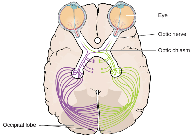
Just below the brain, the optic nerves from each eye meet at the optic chiasm, an X-shaped structure (Figure 3). Here, signals from the right visual field of both eyes are sent to the left hemisphere, and signals from the left visual field are sent to the right hemisphere. This crossing allows both sides of the brain to work together to build a complete visual picture.
Processing in the Brain: The “What” and “Where/How” Pathways
Once visual information reaches the brain, it travels to the occipital lobe at the back of the brain, where sensory input is turned into meaningful perception. The brain processes vision through two main pathways (Milner & Goodale, 2008; Ungerleider & Haxby, 1994):
- The “What” Pathway (Ventral Stream): Identifies what an object is—its color, shape, and meaning.
- The “Where/How” Pathway (Dorsal Stream): Determines where an object is in space and how to interact with it.
For example, when you see a ball rolling across the street, the ventral pathway identifies it as a ball, while the dorsal pathway calculates its movement and location so you can decide whether to catch it or move out of the way.
