How Neurons Communicate
Now that we understand a neuron’s structure, let’s look at how neurons send and receive signals—the process that allows the brain and body to function.
The Resting Potential
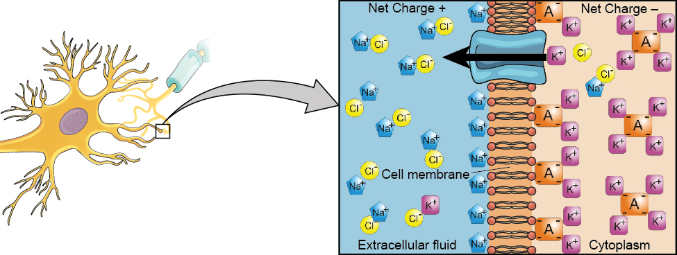
- A neuron’s membrane separates the inside (intracellular fluid) from the outside (extracellular fluid) environment.
- These fluids have different electrical charges, creating an electrical imbalance across the membrane.
- When the neuron is not sending a signal, it is in a resting potential—a state of readiness like a stretched rubber band waiting to release.
- Ions, or electrically charged atoms, line up on either side of the membrane:
- Sodium (Na⁺) ions are more concentrated outside the cell.
- Potassium (K⁺) ions and negatively charged proteins are more concentrated inside the cell.
- This charge difference allows the neuron to respond quickly when activated.
Watch this short video on membrane potential, and why the resting potential of a neuron is negative:
You can view the transcript for “2-Minute Neuroscience: Membrane Potential” here (opens in new window).
Depolarization and the Action Potential
When a neuron receives a signal at its dendrites—usually from neurotransmitters binding to receptors—the following steps occur:
- Gates open in the neuron’s membrane, allowing Na⁺ ions to rush in.
- The inside of the neuron becomes less negative—this is depolarization.
- If depolarization reaches a certain level, the threshold of excitation, the neuron fires an action potential.
The Action Potential
- The action potential is an all-or-none event:
- The neuron either fires at full strength or not at all—there’s no partial firing.
- Once it begins, it propagates down the axon without losing intensity.
- Think of it like sending a text message:
- You can type and rethink it, but nothing happens until you hit “send.”
- Once you do, it delivers completely—you can’t stop it mid-send.
- Because of this property, your brain interprets a stubbed toe as just as “strong” a signal as a touch on your face—the difference lies in which neurons are firing, not how hard they fire.
From Electrical to Chemical Signals
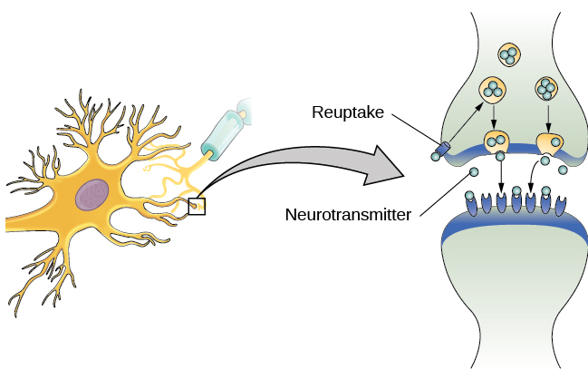
When the action potential reaches the axon terminal:
- Synaptic vesicles release neurotransmitters into the synaptic cleft (the tiny gap between neurons).
- These neurotransmitters cross the cleft and bind to receptors on the next neuron’s dendrites.
- If the signal is strong enough, it triggers another action potential in the receiving neuron—and the process continues.
Reuptake
After a neurotransmitter has done its job:
- Some molecules drift away or are broken down by enzymes.
- Others are reabsorbed into the releasing neuron through reuptake—a recycling process that:
- Clears the synapse for the next signal.
- Helps regulate how much new neurotransmitter the neuron produces.
- This “clean-up” step ensures crisp, on/off signaling in the brain.
Watch this short video to understand how neurons communicate across the synaptic cleft:
You can view the transcript for “2-Minute Neuroscience: Synaptic Transmission” here (opens in new window).