Lobes of the Brain
lobes of the brain
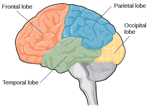
The four lobes of the brain are the frontal, parietal, temporal, and occipital lobes. There are rarely exact locations in the brain responsible for specific functioning, but speaking in general terms, the frontal lobe deals with higher-order thinking and motor movement, the parietal lobe helps process sensory information, the temporal lobes aid in hearing, and the occipital lobes aid in sight.
the frontal lobe
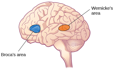
The frontal lobe is located in the forward part of the brain, extending back to a fissure known as the central sulcus. The frontal lobe is involved in reasoning, motor control, emotion, and language. It contains the motor cortex, which is involved in planning and coordinating movement; the prefrontal cortex, which is responsible for higher-level cognitive functioning; and Broca’s area, which is essential for language production.
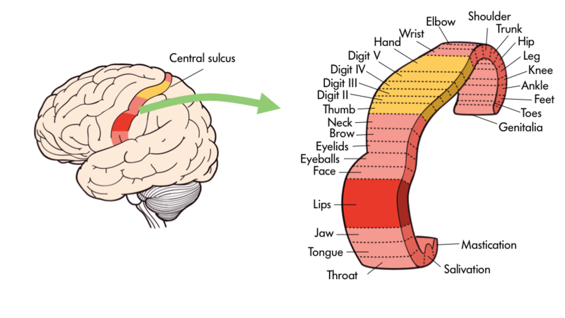
One particularly fascinating area in the frontal lobe is called the “primary motor cortex”. This strip running along the side of the brain is in charge of voluntary movements like waving goodbye, wiggling your eyebrows, and kissing. It is an excellent example of the way that the various regions of the brain are specialized. Interestingly, each of our various body parts has a unique portion of the primary motor cortex devoted to it. Each individual finger has about as much dedicated brain space as your entire leg. Your lips, in turn, require about as much dedicated brain processing as all of your fingers and your hand combined!
the parietal lobe
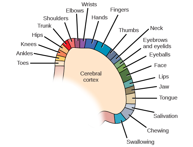
The brain’s parietal lobe is located immediately behind the frontal lobe, and is involved in processing information from the body’s senses. It contains the somatosensory cortex, which is essential for processing sensory information from across the body, such as touch, temperature, and pain. The somatosensory cortex is organized topographically, which means that spatial relationships that exist in the body are generally maintained on the surface of the somatosensory cortex. For example, the portion of the cortex that processes sensory information from the hand is adjacent to the portion that processes information from the wrist.
the temporal lobe
The temporal lobe is located on the side of the head (temporal means “near the temples”), and is associated with hearing, memory, emotion, and some aspects of language. The auditory cortex, the main area responsible for processing auditory information, is located within the temporal lobe. Wernicke’s area, important for speech comprehension, is also located here. Whereas individuals with damage to Broca’s area have difficulty producing language, those with damage to Wernicke’s area can produce sensible language, but they are unable to understand it.
the occipital lobe
The occipital lobe is located at the very back of the brain, and contains the primary visual cortex, which is responsible for interpreting incoming visual information. The occipital cortex is organized retinotopically, which means there is a close relationship between the position of an object in a person’s visual field and the position of that object’s representation on the cortex. You will learn much more about how visual information is processed in the occipital lobe when you study sensation and perception.