Neuron Structure
neuron structure
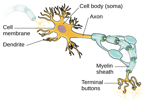
Neurons are the basic building blocks of the nervous system—about 86 billion at birth. Like all cells, they have specialized parts that allow them to process and transmit information.
-
Cell membrane: The neuron’s outer surface is semipermeable, meaning it allows small, uncharged molecules to pass through while blocking larger or charged ones.
-
Soma (cell body): Contains the nucleus, which holds the cell’s genetic material and keeps the neuron functioning.
-
Dendrites: Branching extensions that receive incoming signals from other neurons.
Signal Transmission
-
Axon: A long fiber that carries electrical signals away from the soma. It ends in axon terminals (also called terminal buttons).
-
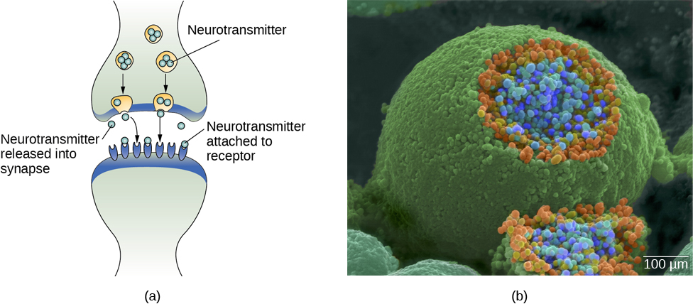
Synaptic vesicles: Found inside the terminals; they store and release neurotransmitters, the chemical messengers of the nervous system.
-
Myelin sheath: A fatty layer produced by glial cells that insulates the axon and speeds up signal transmission.
-
Nodes of Ranvier: Small gaps in the myelin sheath that allow the electrical signal to “jump” from node to node for faster communication.
-
Demyelination: Loss of myelin, as seen in multiple sclerosis (MS), slows neural communication and can cause fatigue, vision problems, and poor coordination.
-
Neural Communication
-
When an electrical impulse reaches the axon terminal, neurotransmitters are released into the synaptic cleft, or gap,—the tiny gap between neurons.
-
These molecules cross the cleft and bind to receptors on the dendrites of the next neuron.
You can view the transcript for “2-Minute Neuroscience: The Neuron” here (opens in new window).